


How Mitochondria Support Fertility and Reproduction
Egg quality and sperm motility are closely tied to mitochondrial health. Learn how to optimize your mitochondria for stronger fertility outcomes.
What to know
Poor mitochondrial function leads to lower egg and sperm quality and challenges with fertility.
As women age, mitochondrial efficiency drops. This decline is a major contributor to age-related fertility decline in women.
Mitochondrial dysfunction is linked to reproductive conditions such as PCOS and endometriosis.
Mitochondrial health plays a key role in sperm quality and motility
Lifestyle changes that target mitochondrial health may be able to improve fertility.
When it comes to baby-making, energy is everything.
Building a human is one of the most demanding processes your body can take on. And that energy comes from your mitochondria. They generate the power your body needs for egg maturation, sperm motility, and fertilization.
Unfortunately, as we age, our mitochondria start to falter, and so does egg quality.
Mitochondrial and Egg Quality
Let’s talk about eggs. Not the ones you scramble, but the ones females are born with!
A single mature egg contains ~100,000 mitochondria, more than any other cell in the human body.[1] When your mitochondria are thriving, your fertility gets a boost.[2] On the other hand, inefficient mitochondria have been linked to infertility.[3]
Age-Related Mitochondrial Decline and Its Impact on Female Fertility
Decline in mitochondrial performance is one of the primary reasons female egg quality declines with age.[4]
As we get older, our mitochondria:[5]
- Accumulate damage
- Produce less energy (ATP)
- Generate more oxidative stress
- Lose efficiency in hormone production
This cellular fatigue is one reason why women in their late 30s and 40s often experience a drop in egg quality and IVF success.[6]
Mitochondrial Health & Reproductive Outcomes
Mitochondria are the site for steroid hormone synthesis, involved in the synthesis and regulation of many different hormones, including estrogen, progesterone, and testosterone. If your mitochondria are underperforming, your hormones likely are too.[7]
Mitochondrial function in eggs is very closely related to fertility outcomes. Certain markers of mitochondrial health (mitochondrial DNA copy number and ATP levels) are associated with higher pregnancy success rates.[8]
On top of that, embryos with mitochondrial deficiencies often fail to implant. The data is so compelling that researchers have proposed using mitochondrial indicators as biomarkers for embryo selection in IVF.[9]
Mitochondrial Health & Reproductive Conditions
Poor mitochondrial function has been associated with conditions impacting reproductive health. For example, studies suggest that eggs in women with endometriosis have a different mitochondrial structure and reduced efficiency, leading to lower fertilization and maturation rates.[10]
In polycystic ovary syndrome (PCOS), mitochondrial function in oocytes is typically impaired, contributing to poor egg quality as well.[11]
Ultimately, the function plays a role in hormone production and balance.
Mitochondria and Sperm Health: Supporting Our Swimmers
Mitochondrial health also plays a role in male fertility. Every sperm cell carries dozens of tiny mitochondria. Research notes that a charged mitochondrial “battery” (normal membrane potential) is essential for human sperm motility and function. If mitochondrial energy output falters, sperm swim poorly, and fertility can suffer.[12]
Sperm Health Matters
Optimizing sperm health is just as important as optimizing egg health! Sperm health can significantly influence pregnancy outcomes. Evidence even suggests that most new genetic mutations originate from the sperm rather than the egg.[13]
Sperm DNA is particularly vulnerable to damage, especially from free radicals and mitochondrial dysfunction.[14] Poor sperm health isn’t just a barrier to conception; it can also impact the health of the pregnancy itself.
Mitochondria Power Sperm Motility and Movement
Efficient mitochondrial function corresponds with plenty of energy for swimming. Sperm with well-charged mitochondria show greater motility and a faster swimming speed.[15]
This link between mitochondria and movement explains why male fertility and sperm energy are tightly connected: any hit to mitochondrial function translates into slow swimming and poor fertility.
Six Biohacks for Mitochondrial Health and Fertility
There are lifestyle changes that can support healthier mitochondria and improved reproductive health.
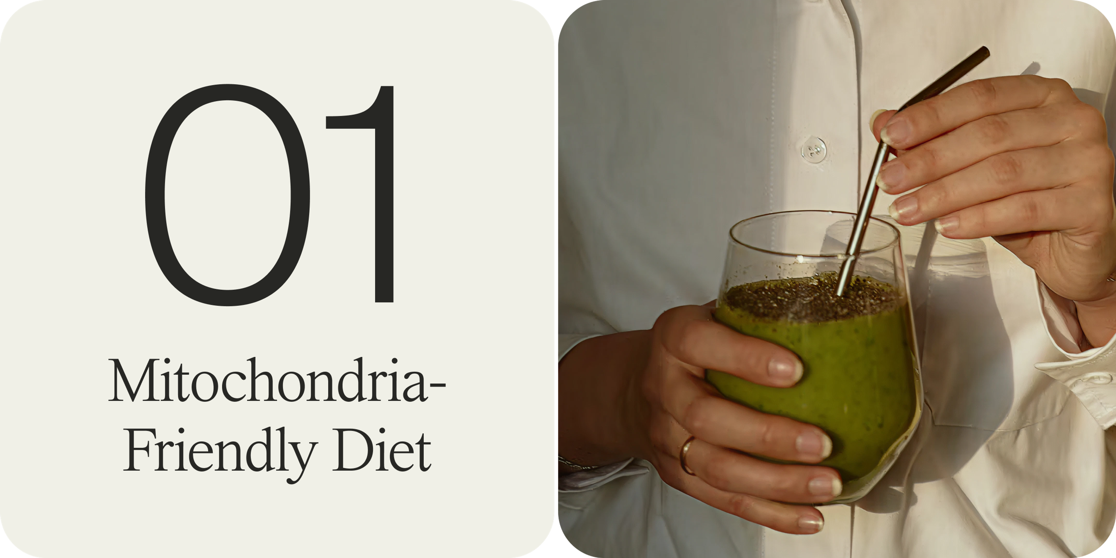
Mitochondria-Friendly Diet
Opt for antioxidant and polyphenol-rich foods[17]. Omega-3s (from fish, walnuts, chia seeds) have also been shown to improve ovarian health.[16] The Mediterranean diet, with its high content of olive oil, nuts, and vegetables, is a great example of a fertility-friendly, mitochondria-supportive eating pattern.

Exercise for Cellular Health
Regular physical activity is one of the best things you can do for your mitochondria. Exercise stimulates mitochondrial biogenesis (the creation of new mitochondria) and improves the efficiency of existing ones.
While the exact mechanism remains unclear, studies suggest that exercise can help with fertility in both men[19] and women[18].

Get Quality Sleep
Never underestimate the power of good sleep for cellular rejuvenation. During deep sleep, our bodies perform critical maintenance, including repairing mitochondrial DNA and removing damaged cell components.
Studies in mice show that chronic sleep deprivation causes oxidative stress and mitochondrial dysfunction in oocytes, leading to fertility decline.
In other words, poor sleep might be aging your eggs[21] and sperm[20].
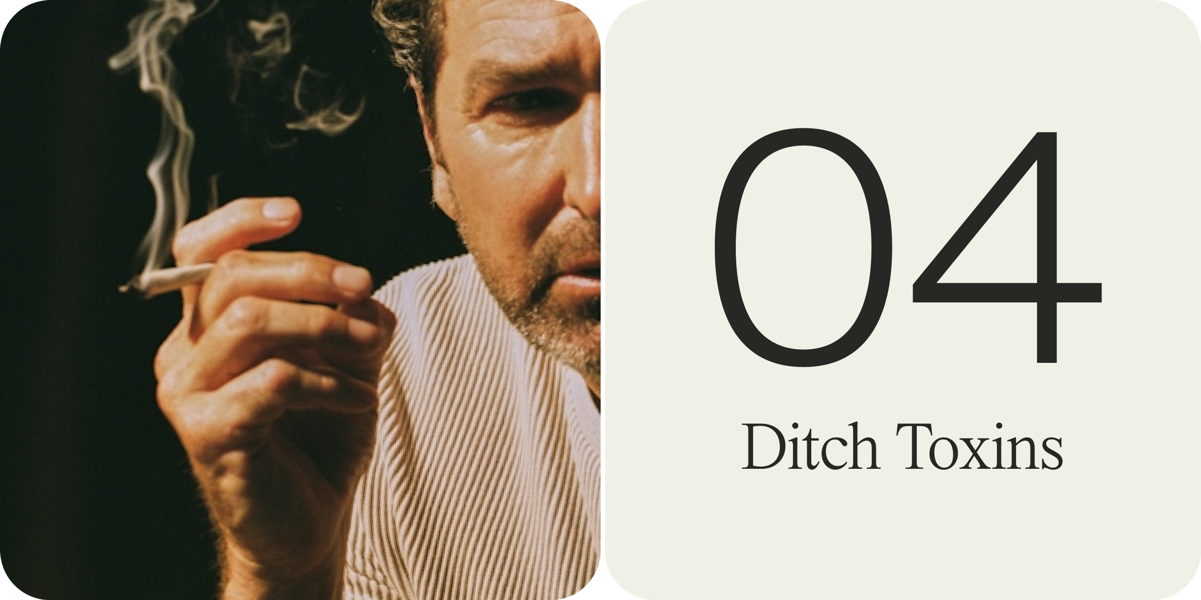
Ditch Toxins
Cigarette smoke, environmental pollutants, and excessive alcohol introduce toxins that generate ROS and directly damage mitochondrial DNA.[24]
Women who smoke tend to experience faster ovarian reserve depletion and earlier menopause.[23] Studies also suggest that smoking can impact mitochondrial function and sperm health in men.[22]
Eliminating smoking and minimizing exposure to toxins (choosing clean beauty/household products, filtering air and water, etc.) will lighten the load on your mitochondria.

Red Light Therapy
Early research highlights the potential of red light therapy to improve biomarkers of mitochondrial health. Animal models find that red light might slow ovarian aging by reducing inflammation and improving mitochondrial function. Similar benefits have been observed in male models, with improvements in sperm mitochondrial health and motility.[25]

Let’s Get (Slightly) Heated
Clinical studies find that mild heat exposure, like using a sauna, has been shown to enhance mitochondrial health in skeletal muscle.[26] While there is no conclusive data on whether saunas can improve mitochondrial signatures in our ovaries, early research is promising.
Urolithin A: A Targeted Solution for Mitochondrial Health
One of the most exciting developments in the world of mitochondrial health is Urolithin A.
While Mitopure has demonstrated benefits in clinical trials regarding mitochondrial function in muscle cells[27], there is currently no clinical research that confirms its impact on fertility or egg quality.
Early, preclinical research in worms indicates that improving mitochondrial recycling could preserve egg quality with age and extend the reproductive span.[28]
Urolithin A is a natural metabolite that is typically produced by our gut when we consume certain polyphenols. It helps your body recycle old, damaged mitochondria through a process called mitophagy. That means more efficient energy production at the cellular level.
The catch? Only 40% of Americans can make any Urolithin A.

Mitopure®: Clinically-Validated Urolithin A
Mitopure is the only clinically validated form of Urolithin A. This highly bioavailable, highly pure form of Urolithin A bypasses gut variability with direct supplementation.
In gold-standard clinical trials, 500mg/day of Mitopure:
- Improved mitochondrial signatures[29]
- Improved cellular energy[30]
- Induced exercise-like effects in muscle cells[31]
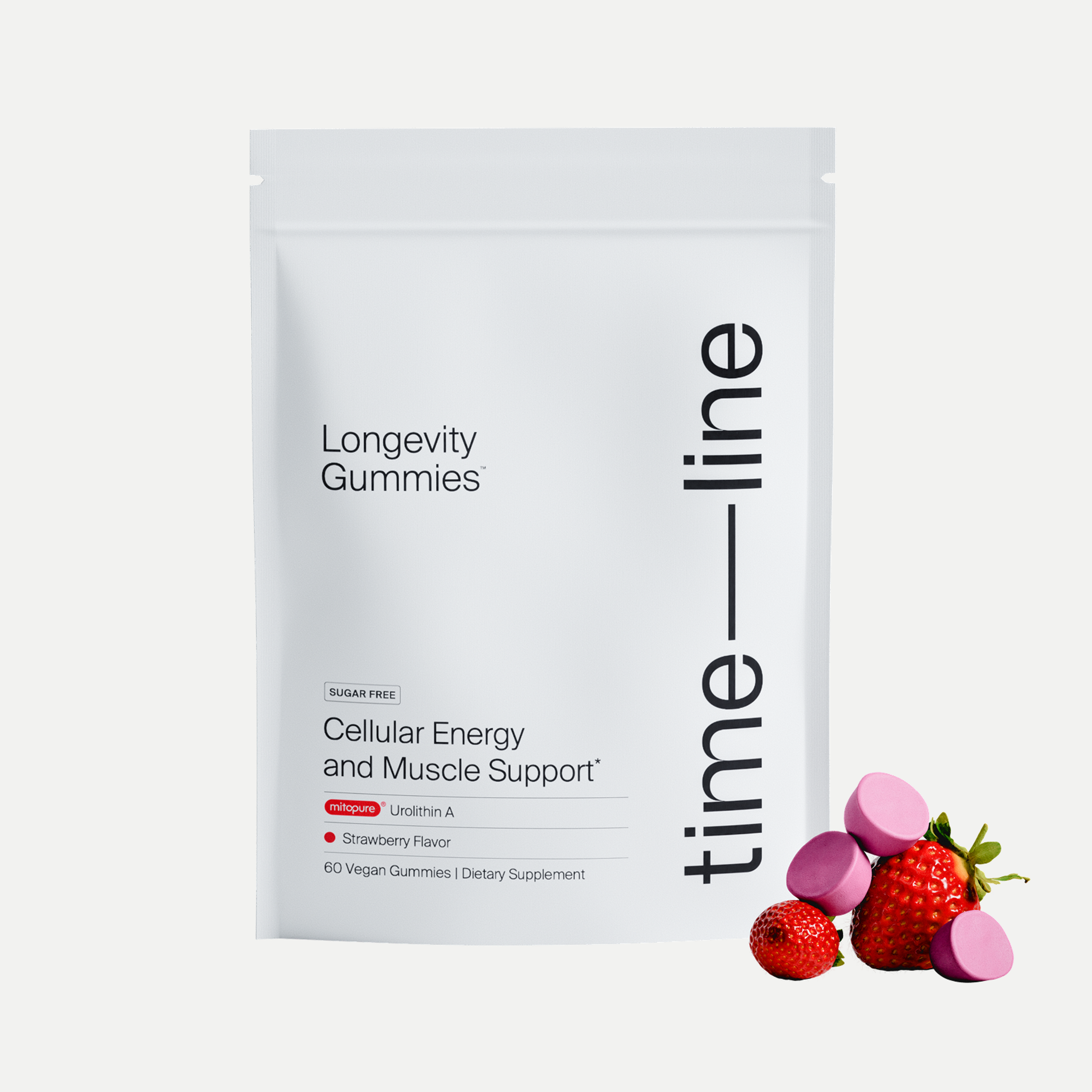
Mitopure Gummies
New4.7 · 424 reviews
A strawberry-flavored dose of cellular energy
Key Takeaways: Mitochondria and Fertility
From egg and sperm quality to hormone production and embryo development, mitochondrial health plays a foundational role in reproductive success for both women and men. As we age, mitochondrial function naturally declines, which can contribute to lower fertility rates and hormone imbalances.
The good news? Mitochondria are highly responsive to lifestyle interventions.
Whether you’re actively trying to conceive or simply want to preserve your reproductive health for the future, investing in your mitochondria is a smart, science-backed strategy.
Authors

Written by
Freelance writer

Reviewed by
Senior Manager of Nutrition Affairs
References
- ↑
Babayev E, Seli E. Oocyte mitochondrial function and reproduction. Curr Opin Obstet Gynecol. 2015 Jun;27(3):175-81. doi: 10.1097/GCO.0000000000000164. PMID: 25719756; PMCID: PMC4590773.
- ↑
Babayev, E., & Seli, E. (2015). Oocyte mitochondrial function and reproduction. Current opinion in obstetrics & gynecology, 27(3), 175–181. https://doi.org/10.1097/GCO.0000000000000164 (https://www.google.com/url?q=https://doi.org/10.1097/GCO.0000000000000164&sa=D&source=docs&ust=1754592073373476&usg=AOvVaw0jZcXlHwdprxZB00kelZO5)
- ↑
Babayev, E., & Seli, E. (2015). Oocyte mitochondrial function and reproduction. Current opinion in obstetrics & gynecology, 27(3), 175–181. https://doi.org/10.1097/GCO.0000000000000164 (https://www.google.com/url?q=https://doi.org/10.1097/GCO.0000000000000164&sa=D&source=docs&ust=1754592073375641&usg=AOvVaw2Tj0buLHqQcsiC3GC7Im_-)
- ↑
Smits, M. A. J., Schomakers, B. V., van Weeghel, M., Wever, E. J. M., Wüst, R. C. I., Dijk, F., Janssens, G. E., Goddijn, M., Mastenbroek, S., Houtkooper, R. H., & Hamer, G. (2023). Human ovarian aging is characterized by oxidative damage and mitochondrial dysfunction. Human reproduction (Oxford, England), 38(11), 2208–2220. https://doi.org/10.1093/humrep/dead177 (https://www.google.com/url?q=https://doi.org/10.1093/humrep/dead177&sa=D&source=docs&ust=1754592073379464&usg=AOvVaw06PCgN_IuiRzxN0VpLNx2L)
- ↑
Smits, M. A. J., Schomakers, B. V., van Weeghel, M., Wever, E. J. M., Wüst, R. C. I., Dijk, F., Janssens, G. E., Goddijn, M., Mastenbroek, S., Houtkooper, R. H., & Hamer, G. (2023). Human ovarian aging is characterized by oxidative damage and mitochondrial dysfunction. Human reproduction (Oxford, England), 38(11), 2208–2220. https://doi.org/10.1093/humrep/dead177 (https://www.google.com/url?q=https://doi.org/10.1093/humrep/dead177&sa=D&source=docs&ust=1755806413857943&usg=AOvVaw2hyaRmlJ3NYE7loajj3rMh)
- ↑
Mobarak, H., Heidarpour, M., Tsai, PS.J. et al. Autologous mitochondrial microinjection; a strategy to improve the oocyte quality and subsequent reproductive outcome during aging. Cell Biosci 9, 95 (2019). https://doi.org/10.1186/s13578-019-0360-5 (https://www.google.com/url?q=https://doi.org/10.1186/s13578-019-0360-5&sa=D&source=docs&ust=1754592073456576&usg=AOvVaw3rJt39qvcapBQQB5md9Diu)
- ↑
Miller W. L. (2013). Steroid hormone synthesis in mitochondria. Molecular and cellular endocrinology, 379(1-2), 62–73. https://doi.org/10.1016/j.mce.2013.04.014 (https://www.google.com/url?q=https://doi.org/10.1016/j.mce.2013.04.014&sa=D&source=docs&ust=1754592073366114&usg=AOvVaw2rKukm1qzQCSJNimtkeaYv)
- ↑
Van Blerkom, J., Davis, P. W., & Lee, J. (1995). ATP content of human oocytes and developmental potential and outcome after in-vitro fertilization and embryo transfer. Human reproduction (Oxford, England), 10(2), 415–424. https://doi.org/10.1093/oxfordjournals.humrep.a135954 (https://www.google.com/url?q=https://doi.org/10.1093/oxfordjournals.humrep.a135954&sa=D&source=docs&ust=1754592073368696&usg=AOvVaw1coKUQ03-C-S9YT1vRBo4d)
- ↑
Seli E. (2016). Mitochondrial DNA as a biomarker for in-vitro fertilization outcome. Current opinion in obstetrics & gynecology, 28(3), 158–163. https://doi.org/10.1097/GCO.0000000000000274 (https://www.google.com/url?q=https://doi.org/10.1097/GCO.0000000000000274&sa=D&source=docs&ust=1754592073401569&usg=AOvVaw2fwSqR9wWFALskaf-UU1XP)
Kim, J., & Seli, E. (2019). Mitochondria as a biomarker for IVF outcome. Reproduction (Cambridge, England), 157(6), R235–R242. https://doi.org/10.1530/REP-18-0580 (https://www.google.com/url?q=https://doi.org/10.1530/REP-18-0580&sa=D&source=docs&ust=1754592073401958&usg=AOvVaw2fPccwEQ-szl3-XfDY7tbn) - ↑
Xu, B., Guo, N., Zhang, X. M., Shi, W., Tong, X. H., Iqbal, F., & Liu, Y. S. (2015). Oocyte quality is decreased in women with minimal or mild endometriosis. Scientific reports, 5, 10779. https://doi.org/10.1038/srep10779 (https://www.google.com/url?q=https://doi.org/10.1038/srep10779&sa=D&source=docs&ust=1754592073405606&usg=AOvVaw2bEqiro8aznDVm5MqwlV79)
- ↑
Xie, C., Lu, H., Zhang, X., An, Z., Chen, T., Yu, W., Wang, S., Shang, D., & Wang, X. (2024). Mitochondrial abnormality in ovarian granulosa cells of patients with polycystic ovary syndrome. Molecular medicine reports, 29(2), 27. https://doi.org/10.3892/mmr.2023.13150 (https://www.google.com/url?q=https://doi.org/10.3892/mmr.2023.13150&sa=D&source=docs&ust=1754592073407786&usg=AOvVaw3p2-C4upvFKZXn1RUs4iRl)
- ↑
Vahedi Raad, M., Firouzabadi, A. M., Tofighi Niaki, M., Henkel, R., & Fesahat, F. (2024). The impact of mitochondrial impairments on sperm function and male fertility: a systematic review. Reproductive biology and endocrinology : RB&E, 22(1), 83.
- ↑
Kong, A., Frigge, M., Masson, G. et al. Rate of de novo mutations and the importance of father’s age to disease risk. Nature 488, 471–475 (2012). https://doi.org/10.1038/nature11396 (https://www.google.com/url?q=https://doi.org/10.1038/nature11396&sa=D&source=docs&ust=1754592073462010&usg=AOvVaw0UNTew4gj7rE9ooqw4JceQ)
- ↑
Fatima, S. (2018). Role of Reactive Oxygen Species in Male Reproduction. . https://doi.org/10.5772/INTECHOPEN.74763 (https://www.google.com/url?q=https://doi.org/10.5772/INTECHOPEN.74763&sa=D&source=docs&ust=1754592073463605&usg=AOvVaw23TC3lT6xp1NdLqzZu7oH_).
- ↑
Costa, J., Braga, P., Rebelo, I., Oliveira, P., & Alves, M. (2023). Mitochondria Quality Control and Male Fertility. Biology, 12. https://doi.org/10.3390/biology12060827 (https://www.google.com/url?q=https://doi.org/10.3390/biology12060827&sa=D&source=docs&ust=1754592073465640&usg=AOvVaw2vwpqclll058eeC-zu6HWE).
- ↑
Nehra, D., Le, H. D., Fallon, E. M., Carlson, S. J., Woods, D., White, Y. A., Pan, A. H., Guo, L., Rodig, S. J., Tilly, J. L., Rueda, B. R., & Puder, M. (2012). Prolonging the female reproductive lifespan and improving egg quality with dietary omega-3 fatty acids. Aging cell, 11(6), 1046–1054. https://doi.org/10.1111/acel.12006 (https://www.google.com/url?q=https://doi.org/10.1111/acel.12006&sa=D&source=docs&ust=1754592073413853&usg=AOvVaw1vdr0nNLI8L91q2Mr49RI-)
- ↑
Ruder, E. H., Hartman, T. J., & Goldman, M. B. (2009). Impact of oxidative stress on female fertility. Current opinion in obstetrics & gynecology, 21(3), 219–222. https://doi.org/10.1097/gco.0b013e32832924ba (https://www.google.com/url?q=https://doi.org/10.1097/gco.0b013e32832924ba&sa=D&source=docs&ust=1754592073410411&usg=AOvVaw1aolO6Kc9Aj0u_0Q9xoFN7)
- ↑
Aria, B., Salegi-Abarghui, A., Lotfi, M., & Mirzaei, M. (2020). Effect of exercise, body mass index, and waist to hip ratio on female fertility. Journal of Basic Research in Medical Sciences, 7, 19-25.
- ↑
Adelowo, O., Akindele, B., Adegbola, C., Oyedokun, P., Akhigbe, T., & Akhigbe, R. (2024). Unraveling the complexity of the impact of physical exercise on male reproductive functions: a review of both sides of a coin. Frontiers in Physiology, 15. https://doi.org/10.3389/fphys.2024.1492771 (https://www.google.com/url?q=https://doi.org/10.3389/fphys.2024.1492771&sa=D&source=docs&ust=1754592073470049&usg=AOvVaw1KNd43XW5JPTqUgM_nrm8w).
- ↑
Du, C., Yang, Y., Chen, J., Feng, L., & Lin, W. (2020). Association Between Sleep Quality and Semen Parameters and Reproductive Hormones: A Cross-Sectional Study in Zhejiang, China. Nature and Science of Sleep, 12, 11 - 18. https://doi.org/10.2147/NSS.S235136 (https://www.google.com/url?q=https://doi.org/10.2147/NSS.S235136&sa=D&source=docs&ust=1754592073471992&usg=AOvVaw1qRDSp5zi1ZQw8JxR_POJT).
- ↑
Yi, Z. Y., Liang, Q. X., Zhou, Q., Yang, L., Meng, Q. R., Li, J., Lin, Y. H., Cao, Y. P., Zhang, C. H., Schatten, H., Qiao, J., & Sun, Q. Y. (2024). Maternal total sleep deprivation causes oxidative stress and mitochondrial dysfunction in oocytes associated with fertility decline in mice. PloS one, 19(10), e0306152. https://doi.org/10.1371/journal.pone.0306152 (https://www.google.com/url?q=https://doi.org/10.1371/journal.pone.0306152&sa=D&source=docs&ust=1754592073425020&usg=AOvVaw3b_vDMaSGXBmEzzElp38rd)
- ↑
Engel, K., Baumann, S., Blaurock, J., Rolle-Kampczyk, U., Schiller, J., Von Bergen, M., & Grunewald, S. (2021). Differences in the sperm metabolomes of smoking and nonsmoking men. Biology of Reproduction, 105, 1484 - 1493. https://doi.org/10.1093/biolre/ioab179 (https://www.google.com/url?q=https://doi.org/10.1093/biolre/ioab179&sa=D&source=docs&ust=1754592073476037&usg=AOvVaw2NosSrwBaaEB08rZCD3M4w).
- ↑
Cui, J., & Wang, Y. (2024). Premature ovarian insufficiency: a review on the role of tobacco smoke, its clinical harm, and treatment. Journal of ovarian research, 17(1), 8. https://doi.org/10.1186/s13048-023-01330-y (https://www.google.com/url?q=https://doi.org/10.1186/s13048-023-01330-y&sa=D&source=docs&ust=1754592073473637&usg=AOvVaw16UXtMroCeN4-f9ZrOrMob)
- ↑
Cui, J., & Wang, Y. (2024). Premature ovarian insufficiency: a review on the role of tobacco smoke, its clinical harm, and treatment. Journal of ovarian research, 17(1), 8. https://doi.org/10.1186/s13048-023-01330-y (https://www.google.com/url?q=https://doi.org/10.1186/s13048-023-01330-y&sa=D&source=docs&ust=1754592073393151&usg=AOvVaw2uL1OzZKN1IyupNI8wYAd1)
Ruder, E. H., Hartman, T. J., & Goldman, M. B. (2009). Impact of oxidative stress on female fertility. Current opinion in obstetrics & gynecology, 21(3), 219–222. https://doi.org/10.1097/gco.0b013e32832924ba (https://www.google.com/url?q=https://doi.org/10.1097/gco.0b013e32832924ba&sa=D&source=docs&ust=1754592073393468&usg=AOvVaw15Js4QvU08VYJiC45AQmwA) - ↑
Catalán, J., Papas, M., Gacem, S., Mateo-Otero, Y., Rodríguez-Gil, J., Miró, J., & Yeste, M. (2020). Red-Light Irradiation of Horse Spermatozoa Increases Mitochondrial Activity and Motility through Changes in the Motile Sperm Subpopulation Structure. Biology, 9. https://doi.org/10.3390/biology9090254 (https://www.google.com/url?q=https://doi.org/10.3390/biology9090254&sa=D&source=docs&ust=1754592073481114&usg=AOvVaw20c1qPSSs_TTMLgc42nZb3).
- ↑
Marchant, E. D., Nelson, W. B., Hyldahl, R. D., Gifford, J. R., & Hancock, C. R. (2023). Passive heat stress induces mitochondrial adaptations in skeletal muscle. International journal of hyperthermia : the official journal of European Society for Hyperthermic Oncology, North American Hyperthermia Group, 40(1), 2205066. https://doi.org/10.1080/02656736.2023.2205066 (https://www.google.com/url?q=https://doi.org/10.1080/02656736.2023.2205066&sa=D&source=docs&ust=1754592073431927&usg=AOvVaw0UuUd6M3X17XtOBB_xkjSX)
- ↑
Andreux, P.A., Blanco-Bose, W., Ryu, D. et al. The mitophagy activator urolithin A is safe and induces a molecular signature of improved mitochondrial and cellular health in humans. Nat Metab 1, 595–603 (2019). https://doi.org/10.1038/s42255-019-0073-4
- ↑
Cota V, Sohrabi S, Kaletsky R, Murphy CT. Oocyte mitophagy is critical for extended reproductive longevity. PLoS Genet. 2022 Sep 20;18(9):e1010400. doi: 10.1371/journal.pgen.1010400. PMID: 36126046; PMCID: PMC9524673.
- ↑
Andreux, P.A., Blanco-Bose, W., Ryu, D. et al. The mitophagy activator urolithin A is safe and induces a molecular signature of improved mitochondrial and cellular health in humans. Nat Metab 1, 595–603 (2019). https://doi.org/10.1038/s42255-019-0073-4
- ↑
Andreux, P.A., Blanco-Bose, W., Ryu, D. et al. The mitophagy activator urolithin A is safe and induces a molecular signature of improved mitochondrial and cellular health in humans. Nat Metab 1, 595–603 (2019). https://doi.org/10.1038/s42255-019-0073-4
- ↑
Singh, Anurag et al. “Urolithin A improves muscle strength, exercise performance, and biomarkers of mitochondrial health in a randomized trial in middle-aged adults.” Cell reports. Medicine vol. 3,5 (2022): 100633. doi:10.1016/j.xcrm.2022.100633