How to Get Rid of Zombie Cells for Longevity
Zombie cells are senescent cells that stop dividing but don't die. Learn how they drive aging, what research shows helps and the role of mitochondria.
What to know
Zombie cells are senescent cells that have permanently stopped dividing but resist dying, and they accumulate in human tissues with age.
Senescent cells release a mix of inflammatory molecules, which links them to age-related inflammation.
Mitochondrial dysfunction and slowed mitophagy help drive cells into senescence, and senescent cells in turn accumulate dysfunctional mitochondria.
In human studies, regular exercise and moderate calorie restriction have both lowered circulating biomarkers of cellular senescence.
Mitopure is clinically proven to trigger mitophagy and improve mitochondrial signatures in the cell.
Aging doesn’t happen all at once. It happens gradually, cell by cell.
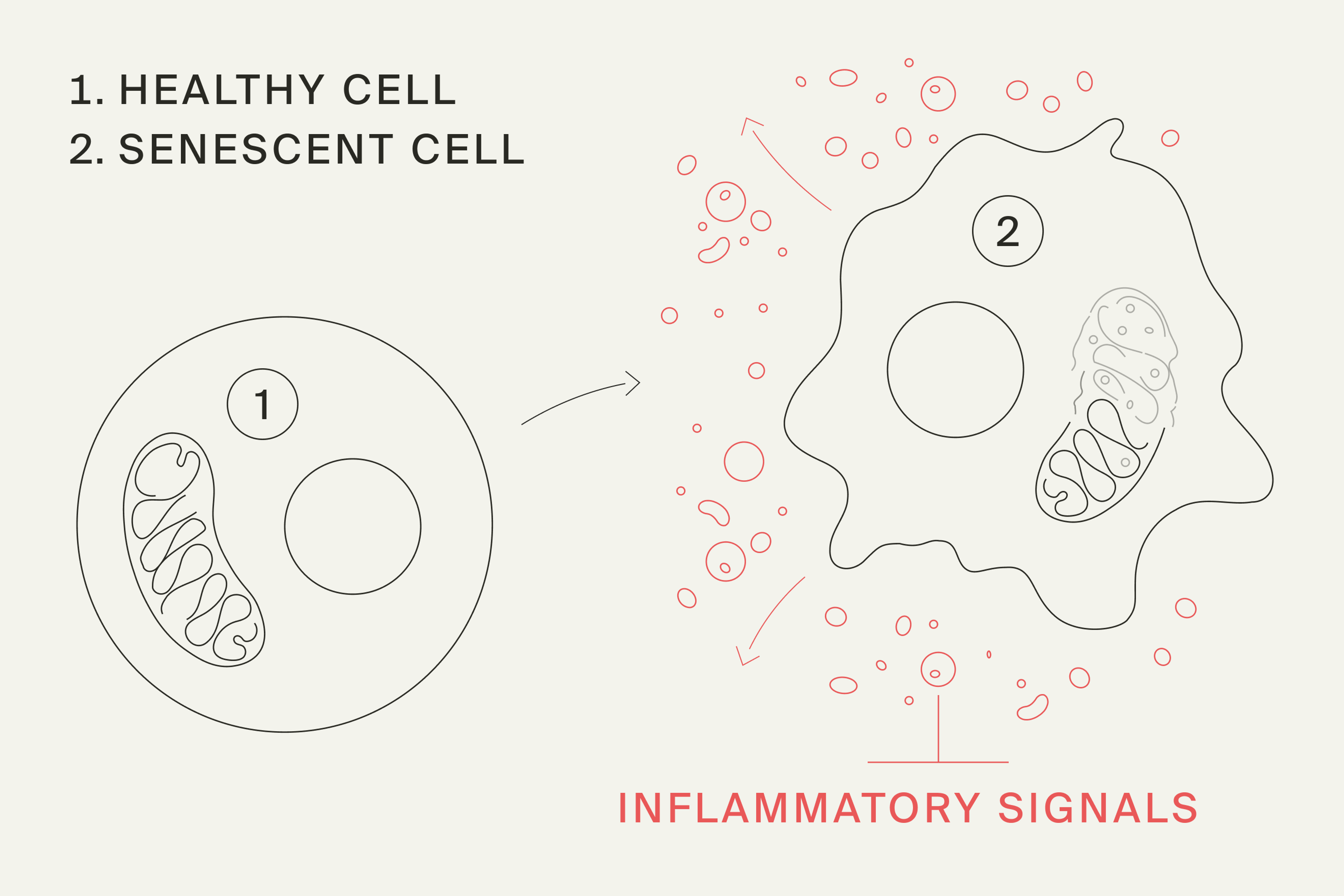
Zombie cells, known scientifically as senescent cells, are damaged or stressed cells that stop dividing but don’t die. Instead, they can linger in tissues and release inflammatory signals that affect the cells around them. As we age, our bodies tend to become less efficient at clearing these cells, allowing them to accumulate over time.
While senescence is a natural process in the body involved in various stages across the lifespan, issues can occur when these cells accumulate.[1]
The accumulation of these “undead” cells doesn’t just give them a haunting name, it can wreak havoc on the body, leading to age-related inflammation and accelerated aging. Understanding them is half the battle. Armed with the right knowledge, foods, supplements, and fasting techniques, we can combat these cellular zombies and pave the way for lasting wellness.
Here’s what we know about why zombie cells accumulate, how your body gets rid of them, and what you can do to support those processes as you age.
What are Zombie Cells?
A zombie cell results from a phenomenon called cellular senescence, a state in which a cell permanently stops dividing but stays alive and metabolically active instead of being cleared away.[2] The nickname comes from that in-between status: not dividing, not functioning normally, but not dead either.
Senescence is not always harmful. In the short term, it acts as a safety mechanism, stopping cells with damaged DNA from multiplying, and it also plays roles in wound healing and embryonic development.[3] Normally, the immune system clears senescent cells once they've done their job.[4]
Problems start when these cells linger. Persistent senescent cells secrete a cocktail of inflammatory proteins, growth factors, and enzymes.[5] These factors can disturb surrounding tissue and even push neighboring cells toward senescence, which is why scientists now list cellular senescence among the hallmarks of aging.[6]
This process is one of the hallmarks of aging, and science is discovering more and more about the role it plays in aging cells and the tools we can use to halt or slow down senescence.[7]
What Creates Zombie Cells?
Cell senescence occurs in response to cellular stressors or damage, ensuring that potentially harmful cells don't continue to divide and proliferate. These cells then cease to multiply and remain alive but dysfunctional.
This is not an ideal scenario, as you want your body to be able to get rid of these damaged cells, not hold onto them. These zombie cells that stick around also release molecules that may trigger age-related inflammation. Mitochondria play a central role in this inflammatory effect. In laboratory studies, cells stripped of their mitochondria lost many senescence features, particularly the inflammatory secretions.[8]
This is why learning how to kill zombie cells or remove senescent cells may help support longevity.
What is the Connection Between Zombie Cells and Aging?
Zombie cells build up with age because the body makes more of them and clears fewer of them as we get older.[9] A systematic review of human tissue studies found that markers of senescence are consistently higher in older tissue samples.[10]
Many age-related processes, such as DNA damage, mitochondrial dysfunction, and disabled mitophagy, are closely tied to the formation of senescent cells.
How Are Zombie Cells and Mitochondria Connected?
Zombie cells and mitochondria are locked in a two-way relationship: mitochondrial dysfunction helps drive cells into senescence, and senescence in turn causes dysfunctional mitochondria to pile up.[11] Mitochondria are the structures inside cells that turn food and oxygen into usable energy, and their decline is itself a hallmark of aging.[12]
Here's how that feedback loop works, according to the research:
- Damaged mitochondria push cells toward senescence: As mitochondria lose efficiency, they produce less energy and more reactive oxygen species, which can damage DNA and trigger senescence.[13]
- Senescent cells fail to recycle mitochondria: Mitophagy, the process that removes worn-out mitochondria, is disrupted in senescent cells, so dysfunctional mitochondria accumulate.[14]
- Dysfunctional mitochondria fuel the inflammation: Mitochondria are required for much of the inflammatory secretory profile that makes senescent cells harmful to surrounding tissue.[15]
Mitophagy also naturally slows with age, which compounds the problem.[16] That's why supporting mitochondrial quality control has become a major focus in aging research. For a deeper look at the process, read our explainer on mitophagy.
Mitopure was developed to support exactly this process. In the first human trial of Urolithin A, older adults who took 500 mg or 1,000 mg daily for 4 weeks showed a molecular signature of improved mitochondrial and cellular health in muscle and blood.[17]

Does Fasting Help Clear Zombie Cells?
Fasting has become a mainstay as a way to improve health because it directly impacts many of the hallmarks of aging. Intermittent fasting activates several biological pathways, including those that can delay senescence.[18]
Additionally, fasting can trigger a cell-recycling process called autophagy, which helps our body remove damaged cells, including senescent cells.[19]
You may be wondering how long you need to fast to kill zombie cells. It is still unclear what the optimal length of time to fast is to stimulate autophagy, but research suggests that at least 24 hours may be needed.[20]
Does Exercise Help Clear Zombie Cells?
Yes, exercise lowers markers of senescence in humans: in older adults, a 12-week structured exercise program reduced circulating biomarkers of cellular senescence.[21] Researchers believe exercise works by boosting autophagy, improving immune clearance of senescent cells, and dampening inflammation.[22]
Exercise is also one of the best-known natural triggers of mitophagy, which ties back to the mitochondria-senescence loop described above.[23]
Do Nutrients and Compounds Help Clear Zombie Cells?
Several plant compounds show activity against senescent cells in lab and animal studies, but none has been proven to remove zombie cells in healthy people.[24] Researchers group these compounds into senolytics, which selectively kill senescent cells, and senomorphics, which calm the age-related inflammation without killing the cell.[25]
Here are some compounds with evidence related to clearing senescence cells:
- Quercetin: This polyphenol is found in fruits and veggies, tea, and wine, and several preclinical trials have shown it has promise as a senolytic compound.[26]
- Resveratrol: Found in grapes and red wine, this natural compound may be able to slow down cell senescence in cells of the blood vessels.[27]
- Curcumin: As an active component of turmeric, curcumin has been studied for its anti-oxidant and anti-inflammatory properties, and there is some evidence to suggest that it may act as a senolytic compound.[28]
- ECGC: The phytochemical epigallocatechin gallate (EGCG) found in green tea may be able to slow cell senescence through the modulation of several pathways involved in this process.[29]
- Urolithin A: Urolithin A is a postbiotic nutrient, meaning our gut microbiome makes it after we ingest polyphenols in foods like pomegranates, berries, and nuts. Preclinical trials suggest that Urolithin A may be able to positively impact cell senescence.[30]
Take Control of Zombie Cells in the Body
You can't switch zombie cells off, but research shows that regular exercise and moderate calorie restriction lower senescence biomarkers in people, and supporting mitochondrial health targets one of the forces that drives cells into senescence.[31] Plant compounds like resveratrol are promising but still largely unproven in healthy humans.[32]
Frequently Asked Questions About Zombie Cells
At what age do zombie cells start building up?
There's no single age when zombie cells start building up: senescence rises gradually with age, and the amount varies widely from one tissue to another. A meta-analysis of 51 studies on human tissue found a consistent positive link between senescence and age, no matter which marker researchers used.[33]
Is there a test for zombie cells?
There's no clinical test for zombie cells yet; measuring senescence is still done only in research settings.[34] Scientists look for a combination of markers, because no single universal marker of senescence exists. Common markers include the protein p16, an enzyme called senescence-associated beta-galactosidase, and SASP proteins circulating in the blood.[35]
What is the difference between senolytics and senomorphics?
Senolytics selectively kill senescent cells, while senomorphics suppress their inflammatory secretions without killing them.[36] Natural senomorphics and senolytics are still mostly being tested in cells and animals.[37]
Can Mitopure help with zombie cells?
Mitopure doesn’t clear zombie cells directly, but it targets one of the mechanisms that drives cells into senescence in the first place. In a human trial, 4 weeks of Urolithin A supplementation produced a molecular signature of improved mitochondrial and cellular health in muscle and blood,[38] and mitochondrial dysfunction is a known trigger for cellular senescence. By activating mitophagy — the process that clears damaged mitochondria — Mitopure supports the same quality-control system that, when it fails, helps push cells toward the senescent state described above.
Authors

Author
Dietitian-Nutritionist, and Health Content Writer

Reviewed by
Lead Regulatory Affairs & Scientific Manager Alliances at Timeline
References
- ↑
Does Cellular Senescence Hold Secrets to Healthier Aging? National Institute on Aging. Published July 13, 2021. Accessed September 29, 2023. https://www.nia.nih.gov/news/does-cellular-senescence-hold-secrets-healthier-aging
- ↑
He, S., & Sharpless, N. E. (2017). Senescence in Health and Disease. Cell, 169(6), 1000–1011. https://doi.org/10.1016/j.cell.2017.05.015
Di Micco, R., Krizhanovsky, V., Baker, D., & d'Adda di Fagagna, F. (2021). Cellular senescence in ageing: from mechanisms to therapeutic opportunities. Nature reviews. Molecular cell biology, 22(2), 75–95. https://doi.org/10.1038/s41580-020-00314-w
- ↑
Ovadya, Y., Landsberger, T., Leins, H., Vadai, E., Gal, H., Biran, A., Yosef, R., Sagiv, A., Agrawal, A., Shapira, A., Windheim, J., Tsoory, M., Schirmbeck, R., Amit, I., Geiger, H., & Krizhanovsky, V. (2018). Impaired immune surveillance accelerates accumulation of senescent cells and aging. Nature communications, 9(1), 5435. https://doi.org/10.1038/s41467-018-07825-3
- ↑
Coppé, J. P., Patil, C. K., Rodier, F., Sun, Y., Muñoz, D. P., Goldstein, J., Nelson, P. S., Desprez, P. Y., & Campisi, J. (2008). Senescence-associated secretory phenotypes reveal cell-nonautonomous functions of oncogenic RAS and the p53 tumor suppressor. PLoS biology, 6(12), 2853–2868. https://doi.org/10.1371/journal.pbio.0060301
- ↑
Di Micco, R., Krizhanovsky, V., Baker, D., & d'Adda di Fagagna, F. (2021). Cellular senescence in ageing: from mechanisms to therapeutic opportunities. Nature reviews. Molecular cell biology, 22(2), 75–95. https://doi.org/10.1038/s41580-020-00314-w
López-Otín, C., Blasco, M. A., Partridge, L., Serrano, M., & Kroemer, G. (2023). Hallmarks of aging: An expanding universe. Cell, 186(2), 243–278. https://doi.org/10.1016/j.cell.2022.11.001
- ↑
López-Otín C, Blasco MA, Partridge L, Serrano M, Kroemer G. Hallmarks of aging: An expanding universe. Cell. 2023 Jan 19;186(2):243-278. doi: 10.1016/j.cell.2022.11.001. Epub 2023 Jan 3. PMID: 36599349.
- ↑
Correia-Melo C, Marques FDM, et al. Mitochondria are required for pro-ageing features of the senescent phenotype. EMBO J. 2016;35(7):724-742. doi: 10.15252/embj.201592862
- ↑
Ovadya, Y., Landsberger, T., Leins, H., Vadai, E., Gal, H., Biran, A., Yosef, R., Sagiv, A., Agrawal, A., Shapira, A., Windheim, J., Tsoory, M., Schirmbeck, R., Amit, I., Geiger, H., & Krizhanovsky, V. (2018). Impaired immune surveillance accelerates accumulation of senescent cells and aging. Nature communications, 9(1), 5435. https://doi.org/10.1038/s41467-018-07825-3
Di Micco, R., Krizhanovsky, V., Baker, D., & d'Adda di Fagagna, F. (2021). Cellular senescence in ageing: from mechanisms to therapeutic opportunities. Nature reviews. Molecular cell biology, 22(2), 75–95. https://doi.org/10.1038/s41580-020-00314-w
- ↑
Tuttle, C. S. L., Waaijer, M. E. C., Slee-Valentijn, M. S., Stijnen, T., Westendorp, R., & Maier, A. B. (2020). Cellular senescence and chronological age in various human tissues: A systematic review and meta-analysis. Aging cell, 19(2), e13083. https://doi.org/10.1111/acel.13083
- ↑
Korolchuk, V. I., Miwa, S., Carroll, B., & von Zglinicki, T. (2017). Mitochondria in Cell Senescence: Is Mitophagy the Weakest Link?. EBioMedicine, 21, 7–13. https://doi.org/10.1016/j.ebiom.2017.03.020
Ghosh-Choudhary, S. K., Liu, J., & Finkel, T. (2021). The role of mitochondria in cellular senescence. FASEB journal : official publication of the Federation of American Societies for Experimental Biology, 35(12), e21991. https://doi.org/10.1096/fj.202101462R
- ↑
López-Otín C, Blasco MA, Partridge L, Serrano M, Kroemer G. Hallmarks of aging: An expanding universe. Cell. 2023 Jan 19;186(2):243-278. doi: 10.1016/j.cell.2022.11.001. Epub 2023 Jan 3. PMID: 36599349.
- ↑
Ghosh-Choudhary, S. K., Liu, J., & Finkel, T. (2021). The role of mitochondria in cellular senescence. FASEB journal : official publication of the Federation of American Societies for Experimental Biology, 35(12), e21991. https://doi.org/10.1096/fj.202101462R
Correia-Melo, C., Marques, F. D., Anderson, R., Hewitt, G., Hewitt, R., Cole, J., Carroll, B. M., Miwa, S., Birch, J., Merz, A., Rushton, M. D., Charles, M., Jurk, D., Tait, S. W., Czapiewski, R., Greaves, L., Nelson, G., Bohlooly-Y, M., Rodriguez-Cuenca, S., Vidal-Puig, A., … Passos, J. F. (2016). Mitochondria are required for pro-ageing features of the senescent phenotype. The EMBO journal, 35(7), 724–742. https://doi.org/10.15252/embj.201592862 - ↑
Korolchuk, V. I., Miwa, S., Carroll, B., & von Zglinicki, T. (2017). Mitochondria in Cell Senescence: Is Mitophagy the Weakest Link?. EBioMedicine, 21, 7–13. https://doi.org/10.1016/j.ebiom.2017.03.020
- ↑
Korolchuk, V. I., Miwa, S., Carroll, B., & von Zglinicki, T. (2017). Mitochondria in Cell Senescence: Is Mitophagy the Weakest Link?. EBioMedicine, 21, 7–13. https://doi.org/10.1016/j.ebiom.2017.03.020
Correia-Melo, C., Marques, F. D., Anderson, R., Hewitt, G., Hewitt, R., Cole, J., Carroll, B. M., Miwa, S., Birch, J., Merz, A., Rushton, M. D., Charles, M., Jurk, D., Tait, S. W., Czapiewski, R., Greaves, L., Nelson, G., Bohlooly-Y, M., Rodriguez-Cuenca, S., Vidal-Puig, A., … Passos, J. F. (2016). Mitochondria are required for pro-ageing features of the senescent phenotype. The EMBO journal, 35(7), 724–742. https://doi.org/10.15252/embj.201592862
- ↑
Sun, N., Yun, J., Liu, J., Malide, D., Liu, C., Rovira, I. I., Holmström, K. M., Fergusson, M. M., Yoo, Y. H., Combs, C. A., & Finkel, T. (2015). Measuring In Vivo Mitophagy. Molecular cell, 60(4), 685–696. https://doi.org/10.1016/j.molcel.2015.10.009
- ↑
Andreux, P. A., Blanco-Bose, W., Ryu, D., Burdet, F., Ibberson, M., Aebischer, P., Auwerx, J., Singh, A., & Rinsch, C. (2019). The mitophagy activator urolithin A is safe and induces a molecular signature of improved mitochondrial and cellular health in humans. Nature Metabolism, 1(6), 595–603. https://doi.org/10.1038/s42255-019-0073-4
- ↑
Liu S, Zeng M, Wan W, Huang M, Li X, Xie Z, Wang S, Cai Y. The Health-Promoting Effects and the Mechanism of Intermittent Fasting. J Diabetes Res. 2023 Mar 3;2023:4038546. doi: 10.1155/2023/4038546. PMID: 36911497; PMCID: PMC10005873.
- ↑
Liu S, Zeng M, Wan W, Huang M, Li X, Xie Z, Wang S, Cai Y. The Health-Promoting Effects and the Mechanism of Intermittent Fasting. J Diabetes Res. 2023 Mar 3;2023:4038546. doi: 10.1155/2023/4038546. PMID: 36911497; PMCID: PMC10005873.
- ↑
Federico Pietrocola, Yohann Demont, Francesca Castoldi, David Enot, Sylvère Durand, Michaela Semeraro, Elisa Elena Baracco, Jonathan Pol, Jose Manuel Bravo-San Pedro, Chloé Bordenave, Sarah Levesque, Juliette Humeau, Alexis Chery, Didier Métivier, Frank Madeo, M. Chiara Maiuri & Guido Kroemer (2017) Metabolic effects of fasting on human and mouse blood in vivo, Autophagy, 13:3, 567-578, DOI: 10.1080/15548627.2016.1271513
- ↑
Englund, D. A., Sakamoto, A. E., Fritsche, C. M., Heeren, A. A., Zhang, X., Kotajarvi, B. R., Lecy, D. R., Yousefzadeh, M. J., Schafer, M. J., White, T. A., Atkinson, E. J., & LeBrasseur, N. K. (2021). Exercise reduces circulating biomarkers of cellular senescence in humans. Aging cell, 20(7), e13415. https://doi.org/10.1111/acel.13415
- ↑
Zhang, X., Englund, D. A., Aversa, Z., Jachim, S. K., White, T. A., & LeBrasseur, N. K. (2022). Exercise Counters the Age-Related Accumulation of Senescent Cells. Exercise and sport sciences reviews, 50(4), 213–221. https://doi.org/10.1249/JES.0000000000000302
Martel, J., Ojcius, D. M., & Young, J. D. (2024). Lifestyle interventions to delay senescence. Biomedical journal, 47(2), 100676. https://doi.org/10.1016/j.bj.2023.100676
- ↑
Zhang, X., Englund, D. A., Aversa, Z., Jachim, S. K., White, T. A., & LeBrasseur, N. K. (2022). Exercise Counters the Age-Related Accumulation of Senescent Cells. Exercise and sport sciences reviews, 50(4), 213–221. https://doi.org/10.1249/JES.0000000000000302
- ↑
Chen, Z., Wu, Y., Lin, Q., Cai, J., Liu, X., & Liang, Y. (2022). Nutrition Interventions of Herbal Compounds on Cellular Senescence. Oxidative medicine and cellular longevity, 2022, 1059257. https://doi.org/10.1155/2022/1059257
- ↑
Di Micco, R., Krizhanovsky, V., Baker, D., & d'Adda di Fagagna, F. (2021). Cellular senescence in ageing: from mechanisms to therapeutic opportunities. Nature reviews. Molecular cell biology, 22(2), 75–95. https://doi.org/10.1038/s41580-020-00314-w
- ↑
Hwang HV, Tran DT, Rebuffatti MN, Li CS, Knowlton AA. Investigation of quercetin and hyperoside as senolytics in adult human endothelial cells. PLoS One. 2018 Jan 9;13(1):e0190374. doi: 10.1371/journal.pone.0190374. PMID: 29315311; PMCID: PMC5760026.
- ↑
Farhadnejad H, Emamat H, Zand H. The Effect of Resveratrol on Cellular Senescence in Normal and Cancer Cells: Focusing on Cancer and Age-Related Diseases. Nutr Cancer. 2019;71(7):1175-1180. doi: 10.1080/01635581.2019.1597907. Epub 2019 Apr 7. PMID: 30955382.
- ↑
Chen Z, Wu Y, Lin Q, Cai J, Liu X, Liang Y. Nutrition Interventions of Herbal Compounds on Cellular Senescence. Oxid Med Cell Longev. 2022 Apr 27;2022:105925
- ↑
Kumar R, Sharma A, Kumari A, Gulati A, Padwad Y, Sharma R. Epigallocatechin gallate suppresses premature senescence of preadipocytes by inhibition of PI3K/Akt/mTOR pathway and induces senescent cell death by regulation of Bax/Bcl-2 pathway. Biogerontology. 2019 Apr;20(2):171-189. doi: 10.1007/s10522-018-9785-1. Epub 2018 Nov 19. PMID: 30456590.
- ↑
Cho, S.I., Jo, ER. & Song, H. Urolithin A attenuates auditory cell senescence by activating mitophagy. Sci Rep 12, 7704 (2022). https://doi.org/10.1038/s41598-022-11894-2
Shi PZ, Wang JW, Wang PC, et al. Urolithin a alleviates oxidative stress-induced senescence in nucleus pulposus-derived mesenchymal stem cells through SIRT1/PGC-1α pathway. World J Stem Cells. 2021;13(12):1928-1946. doi:10.4252/wjsc.v13.i12.1928 - ↑
Englund, D. A., Sakamoto, A. E., Fritsche, C. M., Heeren, A. A., Zhang, X., Kotajarvi, B. R., Lecy, D. R., Yousefzadeh, M. J., Schafer, M. J., White, T. A., Atkinson, E. J., & LeBrasseur, N. K. (2021). Exercise reduces circulating biomarkers of cellular senescence in humans. Aging cell, 20(7), e13415. https://doi.org/10.1111/acel.13415
Aversa, Z., White, T. A., Heeren, A. A., Hulshizer, C. A., Saul, D., Zhang, X., Molina, A. J. A., Redman, L. M., Martin, C. K., Racette, S. B., Huffman, K. M., Bhapkar, M., Khosla, S., Das, S. K., Fielding, R. A., Atkinson, E. J., & LeBrasseur, N. K. (2024). Calorie restriction reduces biomarkers of cellular senescence in humans. Aging cell, 23(2), e14038. https://doi.org/10.1111/acel.14038
Korolchuk, V. I., Miwa, S., Carroll, B., & von Zglinicki, T. (2017). Mitochondria in Cell Senescence: Is Mitophagy the Weakest Link?. EBioMedicine, 21, 7–13. https://doi.org/10.1016/j.ebiom.2017.03.020
- ↑
Yousefzadeh, M. J., Zhu, Y., McGowan, S. J., Angelini, L., Fuhrmann-Stroissnigg, H., Xu, M., Ling, Y. Y., Melos, K. I., Pirtskhalava, T., Inman, C. L., McGuckian, C., Wade, E. A., Kato, J. I., Grassi, D., Wentworth, M., Burd, C. E., Arriaga, E. A., Ladiges, W. L., Tchkonia, T., Kirkland, J. L., … Niedernhofer, L. J. (2018). Fisetin is a senotherapeutic that extends health and lifespan. EBioMedicine, 36, 18–28. https://doi.org/10.1016/j.ebiom.2018.09.015
Hickson, L. J., Langhi Prata, L. G. P., Bobart, S. A., Evans, T. K., Giorgadze, N., Hashmi, S. K., Herrmann, S. M., Jensen, M. D., Jia, Q., Jordan, K. L., Kellogg, T. A., Khosla, S., Koerber, D. M., Lagnado, A. B., Lawson, D. K., LeBrasseur, N. K., Lerman, L. O., McDonald, K. M., McKenzie, T. J., Passos, J. F., … Kirkland, J. L. (2019). Senolytics decrease senescent cells in humans: Preliminary report from a clinical trial of Dasatinib plus Quercetin in individuals with diabetic kidney disease. EBioMedicine, 47, 446–456. https://doi.org/10.1016/j.ebiom.2019.08.069
- ↑
Tuttle, C. S. L., Waaijer, M. E. C., Slee-Valentijn, M. S., Stijnen, T., Westendorp, R., & Maier, A. B. (2020). Cellular senescence and chronological age in various human tissues: A systematic review and meta-analysis. Aging cell, 19(2), e13083. https://doi.org/10.1111/acel.13083
- ↑
Di Micco, R., Krizhanovsky, V., Baker, D., & d'Adda di Fagagna, F. (2021). Cellular senescence in ageing: from mechanisms to therapeutic opportunities. Nature reviews. Molecular cell biology, 22(2), 75–95. https://doi.org/10.1038/s41580-020-00314-w
- ↑
Chen, Z., Wu, Y., Lin, Q., Cai, J., Liu, X., & Liang, Y. (2022). Nutrition Interventions of Herbal Compounds on Cellular Senescence. Oxidative medicine and cellular longevity, 2022, 1059257. https://doi.org/10.1155/2022/1059257
Coppé, J. P., Patil, C. K., Rodier, F., Sun, Y., Muñoz, D. P., Goldstein, J., Nelson, P. S., Desprez, P. Y., & Campisi, J. (2008). Senescence-associated secretory phenotypes reveal cell-nonautonomous functions of oncogenic RAS and the p53 tumor suppressor. PLoS biology, 6(12), 2853–2868. https://doi.org/10.1371/journal.pbio.0060301
- ↑
Di Micco, R., Krizhanovsky, V., Baker, D., & d'Adda di Fagagna, F. (2021). Cellular senescence in ageing: from mechanisms to therapeutic opportunities. Nature reviews. Molecular cell biology, 22(2), 75–95. https://doi.org/10.1038/s41580-020-00314-w
- ↑
Saliev, T., & Singh, P. B. (2025). Targeting Senescence: A Review of Senolytics and Senomorphics in Anti-Aging Interventions. Biomolecules, 15(6), 860. https://doi.org/10.3390/biom15060860
- ↑
Andreux, P. A., Blanco-Bose, W., Ryu, D., Burdet, F., Ibberson, M., Aebischer, P., Auwerx, J., Singh, A., & Rinsch, C. (2019). The mitophagy activator urolithin A is safe and induces a molecular signature of improved mitochondrial and cellular health in humans. Nature Metabolism, 1(6), 595–603. https://doi.org/10.1038/s42255-019-0073-4 (https://doi.org/10.1038/s42255-019-0073-4)
Disclaimer
The information in this article is for informational purposes only and should not be taken as medical advice. Always consult with your medical doctor for personalized medical advice.